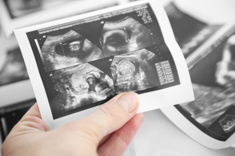
Ultrasound Imaging
A general ultrasound creates ultrasound images that are captured in real time using high frequency sound waves. The images can show the structure and movement of the body’s internal organs, as well as blood flowing through blood vessels. Ultrasound imaging is a noninvasive medical test used to help diagnose and treat medical conditions. Some examples of general ultrasounds are abdominal, thyroid and renal (kidney).
Echocardiogram
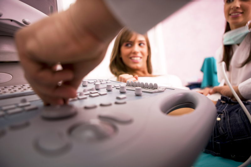
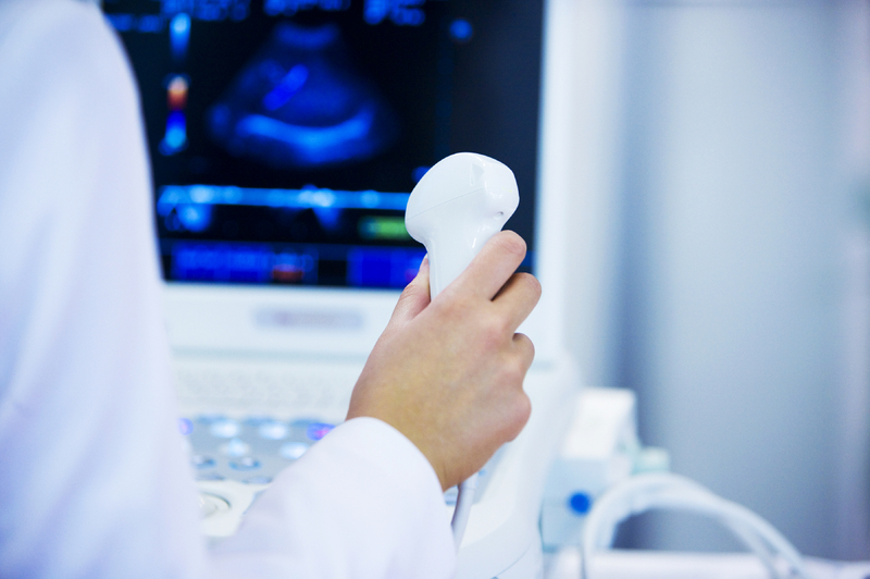
An echocardiogram is a test where ultrasonic (high frequency) sound waves are used to create moving images of the heart. The ultrasonic sound waves are produced by a transducer. They will travel to the heart and 'echo' back, which are then converted to moving images by a computer.
A transthoracic echocardiogram (TTE) is the most commonly used echocardiogram, and involves placing an ultrasound transducer on the chest to produce images.
A transesophageal echocardiogram (TEE) is another type of echocardiogram which is described separately.
A transesophageal echocardiogram or TEE utilizes the insertion of a transducer attached to the tip of a thin tube, or endoscope into the esophagus to visualize the heart, as the esophagus provides optimal viewing. Use of a TEE study provides better and clearer images of the heart and its structures for this reason. TEE can be used as an effective alternative to TTE, if images produced by the TTE are obstructed or unclear due to obesity, bone structure or scarring on the chest wall and certain lung diseases. TEE can be especially useful when evaluating disease of the left upper chamber of the heart (aorta) or valve infection, as these areas are not as clearly visible in a TTE. It is also a useful in detection of abnormal masses inside or outside the heart, identification of clots and causes of strokes and ministrokes, and looking for aortic dissection in a critically ill patient when CT and MRI are not feasible diagnostic options.
A stress echocardiogram also known as ‘Stress echo’ is a simple, painless, non-invasive test used to determine how well your heart and blood vessels are functioning.
Patients with coronary artery disease may not show symptoms at rest. When applied the stress of exercise, the heart pumps more blood. The arteries that are open will dilate to allow more blood but the arteries with blockage will have less blood flow. This difference in the amount of blood flow will produce symptoms like chest pain and discomfort.
The stress echo can either be ‘Exercise stress echo’ or ‘Dobutamine stress echo’.
During exercise stress echo, the patient will be asked to exercise on a treadmill or a stationary bike. The patient will be asked to stop the exercise when the target heart rate for the age is reached or when the patient develops some symptoms that the provider may think are too risky to continue the test. An echocardiogram is done before the exercise(at rest) and later after exercise.
In case of patients who are not able to exercise due to various physical limitations, the stress may be induces by a medication like Dobutamine and the test is then known as 'Dobutamine stress echo'.Dobutamine is adrenaline like substance which when injected mimics exercise and makes the heart beat faster. An echo cardiogram is done before and after the dobutamine injection.
Stress Echo is used to determine:
- how well your heart tolerates any activity
- the progress of known heart valve disease
- to decide the amount of safe exercise before starting a cardiac rehabilitation program after a heart surgery
Vascular Ultrasound
A vascular ultrasound is a noninvasive ultrasound, also known as a duplex that is used to examine the circulation in the blood vessels. A vascular ultrasound can be used to evaluate arteries and veins in nearly any part of the body, including blood vessels in the neck, abdomen, arms and legs. Some examples of vascular ultrasounds are ankle brachial indexes (ABI), radial brachial indexes (RBI), venous and carotid.

Arterial & Venous Duplex
Arterial and venous duplex exam is a very simple, non-invasive and painless test, It incorporates Doppler ultrasound along with traditional ultrasound.
Traditional ultrasound uses sound waves to take images of arteries and veins which will show stenosis(narrowing) and occlusion(blockage) of the arteries and veins. Whereas Doppler ultrasound uses the sound waves to detect the flow of blood in arteries and veins. Being a combination of both the traditional and Doppler ultrasound, duplex exam shows the location, the type and the severity of the vascular disease. Some of the commonly used duplex ultrasounds are carotid duplex, renal duplex, mesenteric duplex along with other vascular ones.
Arterial and venous duplex ultrasound can help diagnose Peripheral Arterial Disease (PAD), Deep vein thrombosis (DVT), Aortic Aneurysm and other vascular disorders. Sometimes it is also used for venous mapping for cardiac bypass surgery.
Vascular Screening
Peripheral artery disease or P.A.D. develops when your arteries become clogged with plaque or fatty deposits that limit blood flow to your lrgs. 1 in every 20 Americans over the age of 50 had P.A.D., a condition that raises the risk for heart attack and stroke. P.A.D. does not always present with symptoms. So it is very important to get screened. At Owensboro Heart and Vascular, we offer 3 simple, non invasive tests for vascular screening-
- Carotid Artery Ultrasound
- ABI- Ankle Brachial Index
- Abdominal Aortic Ultrasound
Click here for a detailed information about vascular screening.
Other Ultrasounds
A general ultrasound creates ultrasound images that are captured in real time using high frequency sound waves. The images can show the structure and movement of the body’s internal organs, as well as blood flowing through blood vessels. Ultrasound imaging is a noninvasive medical test used to help diagnose and treat medical conditions. Some examples of general ultrasounds are abdominal, thyroid and renal (kidney). Owensboro medical practice offers a wide array of ultrasounds including gallbladder, liver, spleen, kidney, thyroid, abdominal, testicular ultrasound, scrotal ultrasound for varicocele, ultrasound for pelvic pain/pelvic congestion.