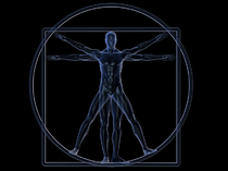
Nuclear Medicine
Nuclear imaging is the branch of imaging involving the administration of trace amounts of radiopharmaceuticals within the body to produce diagnostic images along with the aid of special gamma cameras and computers. These images are used to assess organ function, and is an effective tool for early diagnosis, as the function of the organ is assessed at the molecular level.
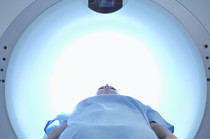
PET CT Scan
PET CT scan is a non-invasive nuclear imaging tool, which is used to assess blood flow to the organs and their function. As with many other nuclear imaging studies, a minute amount of radioactive tracer (Rubidium or 18F-FDG, depending upon the scan) is injected into the blood to assist a Gamma camera and computer with capturing images of the organ. At Owensboro Medical Practice, we offer both Cardiac PET and Oncology PET scans.

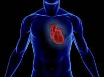
Cardiac PET
PET/CT imaging provides a way to assess the severity of heart disease and measure its impact on heart function. Clinical studies show an important role for PET/CT in screening for coronary heart disease, assessing flow rates and flow reserves, and distinguishing viable from nonviable heart tissue. Myocardial perfusion and myocardial viability are two main clinical applications of a Cardiac PET scan. Read more for detailed information on PET CT imaging available at Owensboro Medical Practice.
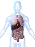
Oncology PET
PET/CT technology has been used effectively for oncology scans. A glucose based radiopharmaceutical known as fludeoxyglucose F18 or FDG is used for oncology PET scans. PET/CT scans show how FDG is taken up by organs and helps the provider decide the location, size and type of tumor. Doctors also use this information to plan the treatment, as well as monitor the effectiveness of treatment.
SPECT Scan
Cardiac SPECT or single photon emission computed tomography also known as myocardial perfusion imaging, is a non-invasive nuclear imaging study used to measure blood flow to the heart at rest and under stress. A radionuclide, such as Technetium is injected into the body, and images are taken with a Gamma camera and computers, which is similar to many other nuclear imaging studies. The blood flow to the heart is measured at rest and after exercise. For exercise, the patient may walk on a treadmill, which is known as a nuclear exercise stress test. If patients are unable to walk along a treadmill, then stress may be induced by a pharmacologic compound, such as Persantine or Adenosine, which causes a simulated response to stress caused by exercise within the body. This is known as a pharmacological nuclear stress test.
A Multi-Gated Acquisition Scan (MUGA) scan, also known as Radionuclide Ventriculography (RNV), Radionuclide Angiography (RNA) or gated blood pool imaging, is a non-invasive nuclear imaging test used to measure the ejection fraction (EF) within your heart. EF is the capacity of the ventricle (lower chamber of your heart) to pump blood with each heartbeat. In this test, a minute amount of radioactive tracer is injected into your vein. A special gamma camera along with the use of a computer creates moving images of the heart that detects size, EF and wall motion of the heart.
A MUGA may be used to detect or monitor Congestive Heart Failure (CHF). Oncologists may use this test to look for pre-existing heart conditions or to estimate the effects of chemotherapy on the heart.
HIDA scan also known as Cholescintigraphy or commonly known as gallbladder scan is a non invasive nuclear imaging tool is used to assess functions of gall bladder and liver.
This procedure uses a radio active tracer (Technetium) which is injected in vein. The tracer is then taken up by bile producing cells of liver from the blood stream and is excreted with bile into the gall bladder via bile duct and eventually into small intestine.
HIDA scan is used to track the flow of bile from liver to gall bladder to small intestineand can help detect gall bladder inflammation(Cholecystitis), bile duct obstruction and congenital problems with bile duct. It can also help assess the progress after liver transplant.