Invasive Testing
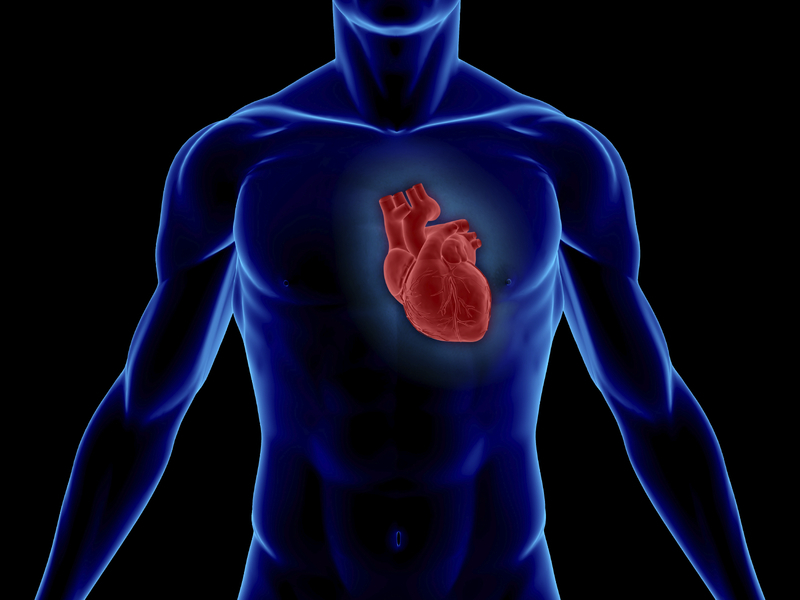
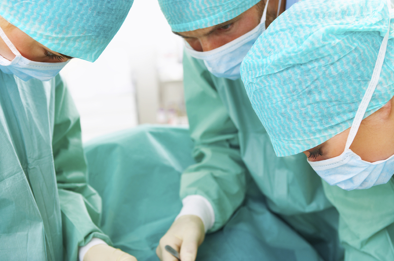
Cardiac catheterization also known as heart or coronary cath, is used to detect or confirm the presence of coronary artery disease, heart valve disease or disease of the aorta. In this procedure, a guide wire is inserted into the blood vessel via the groin area (femoral access) or the wrist area (radial access). A thin tube called catheter is then guided to the heart under x-ray to view the arteries, valves, muscles and aorta. Once the catheter is in place, one or more of the following can be performed: coronary angiography, angioplasty, stent placement, rotablation, LASER atherectomy. Depending on the procedure to be performed, the catheter will have an appropriate attachment.
For more information on cardiac catheterization and related procedures click here.

Coronary Angiography
Carotid Angiography
Intravascular Ultrasound (IVUS), also known as endovascular ultrasound or intravascular echocardiography is an invasive test that uses high frequency sound waves to see the inside of blood vessels. It is performed after cardiac catheterization and sometimes after angioplasty. During this intervention, the catheter with an ultrasound transducer attached to the tip emits sound waves that are converted into images.
IVUS is used to visualize the progression of atherosclerosis within the blood vessels, and to estimate the amount, type and configuration of plaque when traditional angioplasty may not give accurate results. It is also used to determine the type and size of the stent or balloon required, and to evaluate correct placement of the stent. In the aorta, IVUS is used to evaluate plaque buildup and to determine the blood vessels involved in aortic dissection.
TEE
A transesophageal echocardiogram or TEE utilizes the insertion of a transducer attached to the tip of a thin tube, or endoscope into the esophagus to visualize the heart, as the esophagus provides optimal viewing. Use of a TEE study provides better and clearer images of the heart and its structures for this reason. TEE can be used as an effective alternative to TTE, if images produced by the TTE are obstructed or unclear due to obesity, bone structure or scarring on the chest wall and certain lung diseases. TEE can be especially useful when evaluating disease of the left upper chamber of the heart (aorta) or valve infection, as these areas are not as clearly visible in a TTE. It is also a useful in detection of abnormal masses inside or outside the heart, identification of clots and causes of strokes and ministrokes, and looking for aortic dissection in a critically ill patient when CT and MRI are not feasible diagnostic options.
Fractional Flow Reserve
Fractional flow reserve (FFR) is defined as the pressure beyond the coronary stenosis relative to the pressure before the stenosis. After cardiac catheterization FFR is measured to determine need for a coronary intervention when blockages may appear to be borderline severe on angiogram.